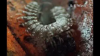
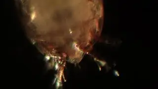
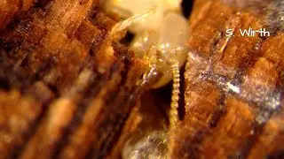
Complex and modified mouthparts in Histiostomatidae mites (4K)
Histiostomatidae possess a bizarre mouthpart apparatus being unique within the Acariformes and representing an amount of characters, which from the phylogenetc point of view can be reconstructed to have evolved in the stem species of that family (so called apomorphies).
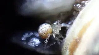
This gnathosoma is a multifunctional organ with the main function to select specific microorganism particles out of their liquid environments.
Recent experiments with a higher videographic resolution and more suitable light conditions than 10 years ago (through-light and up light or one of them depending on the setting) showed that the palpar membrane structures , which more or less surround the entire fore-part (anterior part) of the gnathosoma can act like suckers: When the mite presses its front end of the mouthparts to the underground, an underpressure can be formed based on these membraneous structures. This seemingly facilitates the incorporation of nutrients in that area. I presented such video footage in one of my former mite videos. To get off from the underground requires a jerky upward movement of the whole mouthpart apparatus (also visible in that older video). As I observed different developmental stages of different species, I could conclude that on a smooth surface with randomly dispersed food supply, regular stops and mouthpart-sucking-activities are seemingly a most common behavior of histiostomatids, while a straight forward walking behavior with the gnathosoma permanently touching the ground in order to push microorganism covers to the body`s front side only than occured, when food supply was uniformly dispersed (under artificiall experimental conditions) under uniform moisture conditions.
In my early postdoc-years, still at the FU Berlin, I performed experiments in order to fix mite activities inside their original substrates by filling such a mite-substrate-setting up with 1,1,1,3,3,3-hexamethyldisilazan and warming the corresponding small experimental dish, until the chemical was vaporized. I then sputtered the conserved setting with gold and studied the details on it via scanning-electron-microscopy. It came out that they used the entire laterally bent pedipalpal articles, including the connected palparmembranes, to lean it against the substrate surface, either to stabilize the chelicerae movents or even to support the incorporation of nutrients again by forming a slight underpressure, or both.
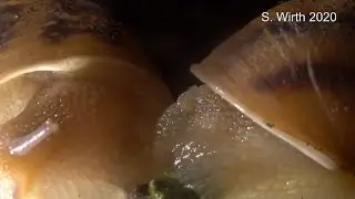
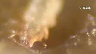
The herewith presented video shows behaviors of a female of the putative new species Histiostoma sp. , which I discovered in beginning of 2019 in sapropel around ponds inside an old gravel pit area in the Berlin forest Grunewald. The footage is presented in slow motion. The question was about how motile the whole gnathosoma apparatus in a histiostomatid species can be and what kinds of movements occured. As the settings, which I in early years of my mite studies used for videographic studies, were simplyfied and thus unnatural (smooth agar surfaces), I thought it being necessary and important to capture behaviors in a complexly sculptured habitat, namely surfaces of decomposing potato pieces (on which most histiostomatid species use to develop well).
It was visible, based on the specimens of my video of this species, that histiostomatid mites can be able to lift up their entire gnathosomas on a sometimes even higher position than the levels of the rest of their bodies. Additionally the gnathosoma can be turned to the right and to the left. Up and down as well as sideward movements of the whole feeding apparatus were often performed and represented obviously flexible reactions of the mite to the surface structure of the substrate and to the availability of suitable nutrients. In this context I was also interested in details of the movements of the chelicera tips themselves.
Although they can be used dagger-like and be accurately inserted into muddy substrate mounts, chelicera tips will also appear in a very fragile and seemingly careful way, when palpating the surface of the substrate underneath. Such chelicera movements are visible in the footage of this video, presented in slow motion (about 25 percent of original speed) and in a digital magnification. I interpret this visible fragility caution of the chelicerae as one option to discover suitable food sources. Other important organs perceive the mite's environment chemically, modified setae, namely the so called solenidia, which might additionally recognize profitable microorganism sources.
Berlin, September 2019
Copyrights Stefan F. Wirth
Watch video Complex and modified mouthparts in Histiostomatidae mites (4K) online, duration hours minute second in high quality that is uploaded to the channel Stefan F. Wirth 22 September 2019. Share the link to the video on social media so that your subscribers and friends will also watch this video. This video clip has been viewed 162 times and liked it 4 visitors.