Structure of Inner Ear
Structure of Ear : • Structure of Ear | Flow Charts and An...
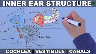
The inner ear sits within the temporal bone in a complex cavity called the bony labyrinth. A central area known as the vestibule contains two small fluid-filled recesses, the utricle and saccule. These connect to the semicircular canals and the cochlea. There are three semicircular canals angled at right angles to each other which are responsible for dynamic balance. The cochlea is a spiral shell-shaped organ responsible for the sense of hearing. These structures together create the membranous labyrinth.
The bony labyrinth refers to the bony compartment which contains the membranous labyrinth, contained within the temporal bone. The inner ear structurally begins at the oval window, which receives vibrations from the incus of the middle ear. Vibrations are transmitted into the inner ear into a fluid called endolymph, which fills the membranous labyrinth. The endolymph is situated in two vestibules, the utricle and saccule, and eventually transmits to the cochlea, a spiral-shaped structure. The cochlea consists of three fluid-filled spaces: the vestibular duct, the cochlear duct, and the tympanic duct.[7] Hair cells responsible for transduction—changing mechanical changes into electrical stimuli are present in the organ of Corti in the cochlea
Watch video Structure of Inner Ear online, duration hours minute second in high quality that is uploaded to the channel Hussain Biology 01 January 1970. Share the link to the video on social media so that your subscribers and friends will also watch this video. This video clip has been viewed 1,042 times and liked it 40 visitors.