Swelling Image | Microfluidic systems for treatment screening under controlled hypoxia
Tumour hypoxia (low oxygen levels) can impact the effectiveness of cancer treatments. Abnormal blood vessels in tumours can lead to oxygen gradients and chronic and transient (intermittent) hypoxic regions, with hypoxia/reperfusion rates of a few cycles per hour. Transient hypoxia can promote tumour aggressiveness and changes in cell phenotype compared with chronic hypoxia. Including these effects in early in vitro screening stages could yield results more predictive during subsequent in vivo tests. Since state-of-the-art in vitro platforms such as well plates, glass dishes, hypoxia chambers, and even custom stirred hypoxic vessels can’t easily reproduce these effects due to long equilibration times (hours), we use microfluidic devices which offer the potential to control and reproduce more realistic environments due to their smaller size scales.
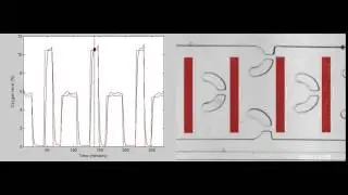
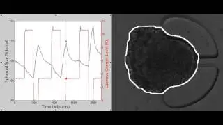
Video showing the response of an MCF-7 breast cancer tumour spheroid to cyclic hypoxia within a microfluidic oxygen control device. Left: Spheroid size (black) and oxygen level supplied to the control device (red) during exposure to a cycling oxygen profile (cycling between 0%, 3%, and 10% oxygen). Right: Brightfield video of spheroid overlaid with the segmentation (white line) used to calculate the spheroid size plotted on the left. The spheroid increases in size when exposed to 0% oxygen and decreases again when exposed to 3% and 10% oxygen, showing evidence of dynamic swelling and shrinkage during cycling hypoxia.
Watch video Swelling Image | Microfluidic systems for treatment screening under controlled hypoxia online, duration hours minute second in high quality that is uploaded to the channel UBC Electrical and Computer Engineering 27 July 2016. Share the link to the video on social media so that your subscribers and friends will also watch this video. This video clip has been viewed 223 times and liked it 0 visitors.